Medical Imaging, Explained
- Category: Choose health
- Posted On:

X-ray, CT scans and MRI exams are three of the most common tools used in medical imaging. Here’s a rundown of these tests and how they’re used.
X-RAY
X-ray exams allow radiologists to take internal pictures of your body using small doses of radiation. This test is often used to confirm bone fractures but may also be used to diagnose pneumonia, blocked intestines and other illnesses. Two types of X-ray tests include mammograms, which detect breast cancer, and dual-energy X-ray absorptiometry, which measures bone mineral density and can reveal osteoporosis.
CT SCANS
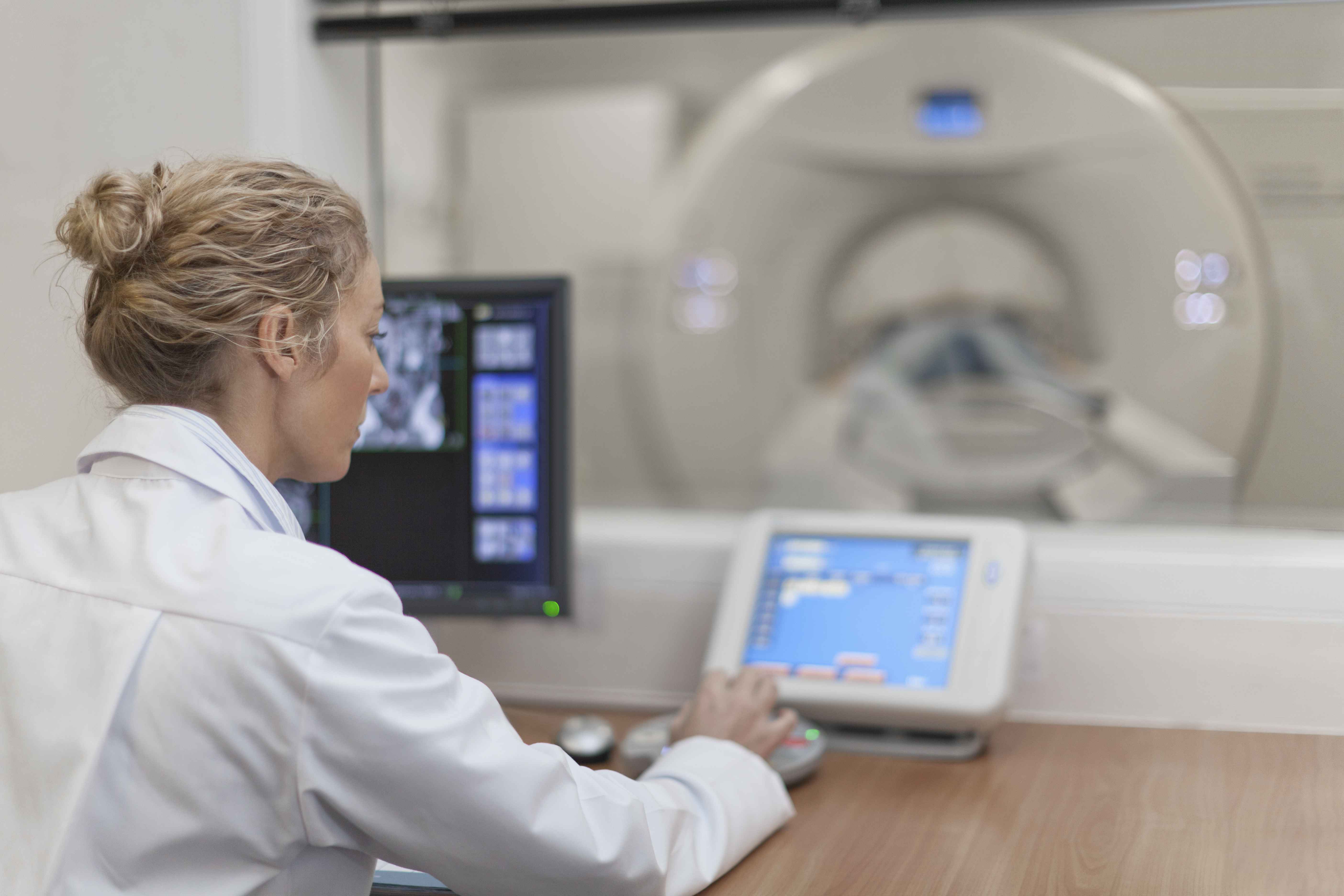
CT, or computed tomography, scans combine X-ray with computer technology to create detailed, tow-or three-dimensional pictures of the body. During a CT scan, you lie still on a table eas the machine encircles you to take pictures in sections. Computers then reconstruct these images into a multi-dimensional model of your body. CT scans are often used to gain a better view of soft tissues, such as those in the chest and stomach.
MRI
MRI, or magnetic resonance imaging, employs large magnets and radio frequencies, instead of radiation, to create images in greater detail and clarity than other imaging tests can. MRI can help doctors diagnose or better understand a wide variety of health issues that affect your brain, heart, skeleton and virtually any other part of your body. For example, MRI can help radiologists better see abnormalities detected on a mammogram or understand how a strike has affected the brain.
Archbold offers advanced imaging services, including CT, MRI and 3D mammography. If you have questions, email rajense@archbold.org.


